
They say about 80% of the information we take in comes through the eyes - such an invaluable part of the human body. However as the years pass, our eyes become more susceptible to different kinds of disorders, and if treatment is too late, they can result in loss of vision. As soon as you have symptoms that concern you, it's best to visit a specialist as soon as possible. Not all eye diseases have clear symptoms for early detection, so if you're unsure or uneasy about your eyes' condition, don't hesitate to stop by for an appointment.
|
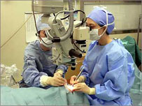
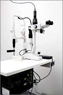
Surgical Environment
|
|
 Cataract Cataract
Do you see double, feel like you're looking through a mist, or feel sensitive to bright lights? These could be early symptoms of a cataract developing. A cataract is when an opacity forms on the lens of the eye, which prevents light from fully reaching the retina. This can cause a haziness and loss of vision. The most common cause of cataracts is aging. It is said that 60% of individuals in their 60's and 80% in their 70's suffer from age-related cataracts. Other causes of cataracts include external injuries to the eye, diabetes, and atopic dermatitis. Despite the differing causes, the result of a gradual opacity of the lens is a common symptom in all forms of cataracts.
What should I do if I am suffering from a cataract?
There are 2 treatment methods: one using eye drops, and one involving surgery.
Eye drops can be effective in preventing the oxidization of proteins and sugars in the lens, so they are an effective treatment in light cataract cases where vision has not yet been affected. However since eye drops cannot fully prevent the progress of cataracts, regular eye checkups will be necessary.
As it's unlikely that eye drops will be effective in returning eyesight once it has been lost, in cases where the eyesight has already been affected, surgery is recommended for dealing with cataracts. In cases where surgery is needed, Oji Eye Clinic's director, Dr. Yamamoto, performs eye surgery at Komagome Hospital. The operation consists of removing the lens that has been affected by the cataract and replacing it with a new synthetic lens. In most cases it is a relatively quick operation only requiring a local anesthesia (in eye drop form). The affected lens is emulsiphied with ultrasonic waves and removed while leaving the majority of the lens capsule behind.
The most recent method of cataract surgery is called "Small Incision Cataract Surgery". A typical lens is 6mm; however, in small incision cataract surgery, new soft materials are used to make the lens so that it can be folded in half or in thirds before insertion. This results in a much smaller incision and less damage to the eyeball from the surgery. Generally the incision is made just above the cornea; however, in cases of astigmatism and other causes, incisions are made toward the ear side or from the top in order to prevent creating any new astigmatism in the eye. Also existing astigmatism can be correct at that time with an astigmatism correcting lens. With "small incision cataract surgery," the incision is small enough that there is rarely need for stitching back the incision, which results in almost no inflammation to the eye. This way the operation is much safer and can be done in one day with no overnight stay at the hospital necessary. Of course depending on the conditions, sometimes hospital admission can be necessary. The recovery time is different in every case, but in general, from the next day to within one week of the operation, eyesight should begin to improve.
Secondary Cataract
A few months or a few years after cataract surgery, the eye will sometimes once again begin to haze up and become sensitive. This is called a "secondary cataract", and it is caused by the posterior lens capsule fogging up. With secondary cataracts, an operation is not necessary, and it can be quickly cleared up with laser treatment. Eyesight will quickly recover and no hospital admission is necessary. |
|
|
|
|
|
  |
|
Left image: Cataract
Right image: After cataract surgery, synthetic lens
※Click on the image to see a larger picture in a separate window |
|
|
|
 |
|
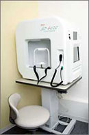
Field of Vision Gauge |
|
Glaucoma
Glaucoma is a disease when something damages the optical nerve causing field of vision to narrow, mainly caused by an increase in intraocular pressure. It's said that about 1 in 17 people over 40 and 1 in 7 people over 70 suffer from glaucoma.
There are almost no obvious early symptoms of glaucoma, so usually you become aware of it after it has already progressed. It can be detected early with ophthalmotonometry, funduscopy, of field of vision tests. There are a few types of glaucoma. They're organized into 3 broad categories: primary glaucoma (open-angle or closed-angle), congenital glaucoma, and secondary glaucoma.
Although there is currently no complete cure for glaucoma, the most effective treatments control ocular tension and halt the advance of the illness. The 3 methods of treatment are through medicine, laser treatment, and surgery. The medicines that are used can reduce the production of or increase discharge of aqueous humor (the fluid that forms the shape of the eyeball). At first you start with one kind of eye drop and, while observing the condition of the eyes, the treatment can change to another kind of eye drop or a combination of a few kinds. Also in cases where eye drops are insufficient, internal medicines can also be used. At Oji Eye Clinic, a laser operation can be done to open a hole in the iris, or using a laser on the trabeculum, production of aqueous humor can be induced. In cases where glaucoma causes the lens to swell or refractory cases of glaucoma, more complex operations at Komagome Hospital are also used.
|
|
|
|
|
|
Laser Ophthalmoscope |
|
Diabetes, High Blood Pressure, and other Lifestyle Disorders
In Japan 7,400,000 people are at risk of diabetes, and the number of people who fall victim to visual disabilities caused by diabetes has risen to 3000 per year. Including blindness, diabetic retinopathy has become one of the leading causes of visual disorders.
At the far end of the eye, on the fundus there is a nerve membrane called the retina, which is covered by many capillaries. Arterialsclerosis causes blood vessels in patients with high blood pressure or high blood fat to narrow. In the blood vessels of diabetes patients there is a high level of sugar which can easily clot causing the capillaries in the retina to decrease or cause a burden on the vascular wall causing hemorrhage in the fundus of the eye. That causes the blood flow to decrease and deprive the retina from necessary oxygen and nutrients, which becomes a cause of increased diabetic retinopathy. In cases where diabetic retinopathy advances, new blood vessels are formed causing bleeding in the vitreous body of the eye, traction retinal detachment, and in conjunction with rubeotic glaucoma, can lead to blindness. Because it's impossible to detect early stages of membrane damage diabetes, regular tests at an eye clinic are very important.
Blood sugar control is essential in the treatment of diabetic retinopathy. Blood sugar control alone can sometimes improve hemorrhaging in the eye in cases of simple retinopathy. Optimal treatment is recommended in collaboration with the patient's family practitioner.
In cases of advanced diabetic retinopathy laser coagulation becomes necessary. With laser treatment the formation of new blood vessels can be avoided and bleeding and white spots can be cured. This is an outpatient procedure, and, depending on the level of advancement, can be divided and carried out in multiple stages. Laser coagulation cannot restore lost vision, but it can hinder the advancement of the retinopathy to a certain extent.
In cases where new blood vessels rupture and cause bleeding in the vitreous body or retinal detachment, admission to the hospital and surgery are required.
|
|
|
|
|
|
  |
|
Left image: Diabetic Retinopathy
Right Image: Diabetic Retinopathy after laser treatment
※Click on the image to see a larger picture in a separate window |
|
|
|
|
|
Laser Ophthalmoscope |
|
Myopia/Myodesopsia (a bug-like shadow at the front of the eye)
Myodesopsia is mostly caused by old age, but to check if the cause is physiological or caused by disease, funduscopy is recommended.
A thin retina in patients with myopia leads to vitreous detachment caused by age or other origins, which causes a retinal tear. From the center of that retinal tear, the retina becomes detached from the lower layer and can float into the vitreous body. The initial symptom of this sort of phenomenon is a sudden increase in the floating bodies over the eye. If it's left untreated, this will eventually lead to blindness.
The treatment of a retinal tear uses a laser to harden the area surrounding the tear (light coagulation method) preventing retinal detachment. This is an outpatient operation; however, if the retina detaches, hospitalization and surgery will become necessary.
Also diabetes, high blood pressure, external injuries causing hemorrhage in the fundus, alergic reactions to bacteria or viruses, or inflammation of the uvea can cause the symptoms of myodesopsia.
Treatment as early as possible is essential, so please don't hesitate to consult us. |
|
|
|
|
|
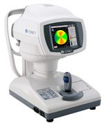
Cornea Topography
|
|
Conditions that can't be corrected with glasses
We use a cornea topography to detect the early stages of conical cornea and irregular astigmatism that can't be observed with a usual slit-lamp and consult about how to prevent against advancement, and we can provide pertinent contact lenses in such cases.
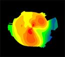 Conical Cornea color code map Conical Cornea color code map |
|
|
|
|
|
|
|
Inverted Eyelash, Slack Eyelids
Mild cases of inverted eyelids can be treated with simple removal, but in serious cases it can cause corneal disorders; therefore, it must be removed by surgery. The hospital director performs a "Dermabrasion Double Eyelid Surgery" to correct the problem.
|
|
|
|
|
|
|
|
Watery Eye, Dry Eye
We use noninvasive, quantitative tools for precise diagnosis of the tear fluid turnover and function of the corneal epithelium barrier, and for serious cases of dry eye, we use a treatment that blocks the exit of tears with a lacrimal opening plug allowing tears to accumulate or perform plastic surgery on the outer surface of the eye. |
|
|
|
|
|
|
|
Congenital Nasolacrimal Duct Obstruction
Immediately after birth, eye mucus and lacrimation are observed. Tears are sucked up from the lacrimal points located in the medial angle of the eye and flow between the lacrimal duct, lacrimal sac and nasal cavity. The cause of this is incomplete development of the nasal cavity in the nasolacrimal duct. The treatment begins with a massage of the lacrimal sac with antibiotic tear drops. If symptoms don't improve more than 3 months after birth, in order to wash the lacrimal passage, a slender wire (bougie) is passed through it. |
|
|
|
|
 |
 |
|
|